Scanning a Doberman for a Compressed Neck
There's nothing quite as heartbreaking as seeing your normally agile Doberman start to falter, perhaps taking an unsteady step, dragging a back paw, or holding their head low as if carrying the weight of the world. These subtle, and sometimes not-so-subtle, changes can be incredibly alarming, and for many Doberman owners, they immediately bring to mind one of the breed's most challenging health issues: Wobbler Syndrome, or cervical spondylomyelopathy. This guide is here to walk you through the essential diagnostic steps your veterinarian will take to scan your Doberman for a compressed neck, explaining each procedure so you can navigate this worrying time with confidence and understanding.
The Worrying Signs of a Compressed Doberman Neck
It’s easy to dismiss early signs as just an awkward moment or a bit of stiffness after a long nap, but with a Doberman, these seemingly minor issues can quickly snowball into something far more serious. Recognizing the nuances of how a compressed neck can present in our powerful, elegant companions is the first step toward getting them the help they need.
Decoding the Doberman Wobble: Early Symptoms
The nickname "Wobbler Syndrome" isn't an exaggeration; it vividly describes the hallmark symptom of this condition. Often, owners first notice a change in their Doberman's gait, particularly in the hind limbs. Your dog might appear uncoordinated, almost drunk, struggling to place their feet properly. Imagine your Doberman usually trotting confidently, then suddenly they start to sway or scuff their back paws on the ground, wearing down the tops of their nails. This can be a subtle sign of proprioceptive deficits, meaning they aren't fully aware of where their limbs are in space.
You might also observe a reluctance to move their head or neck, perhaps refusing to look up at you for a treat, or simply holding their head in a fixed, lowered position. This can be a clear indicator of neck pain or stiffness. Sometimes, they might even yelp when turning their head or when you gently touch their neck area. These early indicators are crucial and shouldn't be ignored. Even a slight change in their normally fluid movement, or a new hesitancy when jumping or going up stairs, warrants a closer look. If you've noticed A Doberman That Wobbles on Its Back Legs, it's time to talk to your vet.
Progression of Neurological Symptoms
Left undiagnosed or untreated, these early signs often worsen and can involve the front limbs as well. As the spinal cord compression becomes more severe, the neurological deficits become more pronounced. What started as a slight wobble might turn into overt ataxia, where your Doberman struggles significantly to walk in a straight line, stumbles frequently, or even falls. Muscle weakness and atrophy, especially in the shoulders, can become noticeable. Their strong, muscular Doberman physique might start to diminish in certain areas.
In some severe cases, Dobermans can experience acute episodes of extreme pain, hind limb paralysis, or even sudden collapse. This progression can be frighteningly quick or agonizingly slow, unfolding over months or even years. The insidious nature of Wobbler Syndrome is that the Age Doberman Wobbler Signs Appear can vary widely, from relatively young adulthood to senior years, making vigilance throughout your Doberman's life essential. The impact on their quality of life can be profound, turning an active, joyful dog into one that struggles with everyday movements.
Initial Steps: What Your Vet Looks For First
When you bring your Doberman to the vet with concerns about a compressed neck, the diagnostic process begins with a thorough and systematic approach. It’s not just about looking at the neck; it’s about understanding the entire neurological picture.
The Neurological Examination: More Than Just a Check-Up
Your veterinarian, or ideally a veterinary neurologist if one is available, will perform a comprehensive neurological examination. This is far more in-depth than a standard wellness check. They'll assess your Doberman's gait, observing them walking, trotting, and turning in various ways. They’ll look for any signs of ataxia, weakness, or scuffing. They’ll test their proprioception by flipping their paws over and seeing how quickly your Doberman corrects them. They'll check reflexes in all four limbs, evaluate muscle tone, and assess for any signs of pain or discomfort when manipulating the neck and spine.
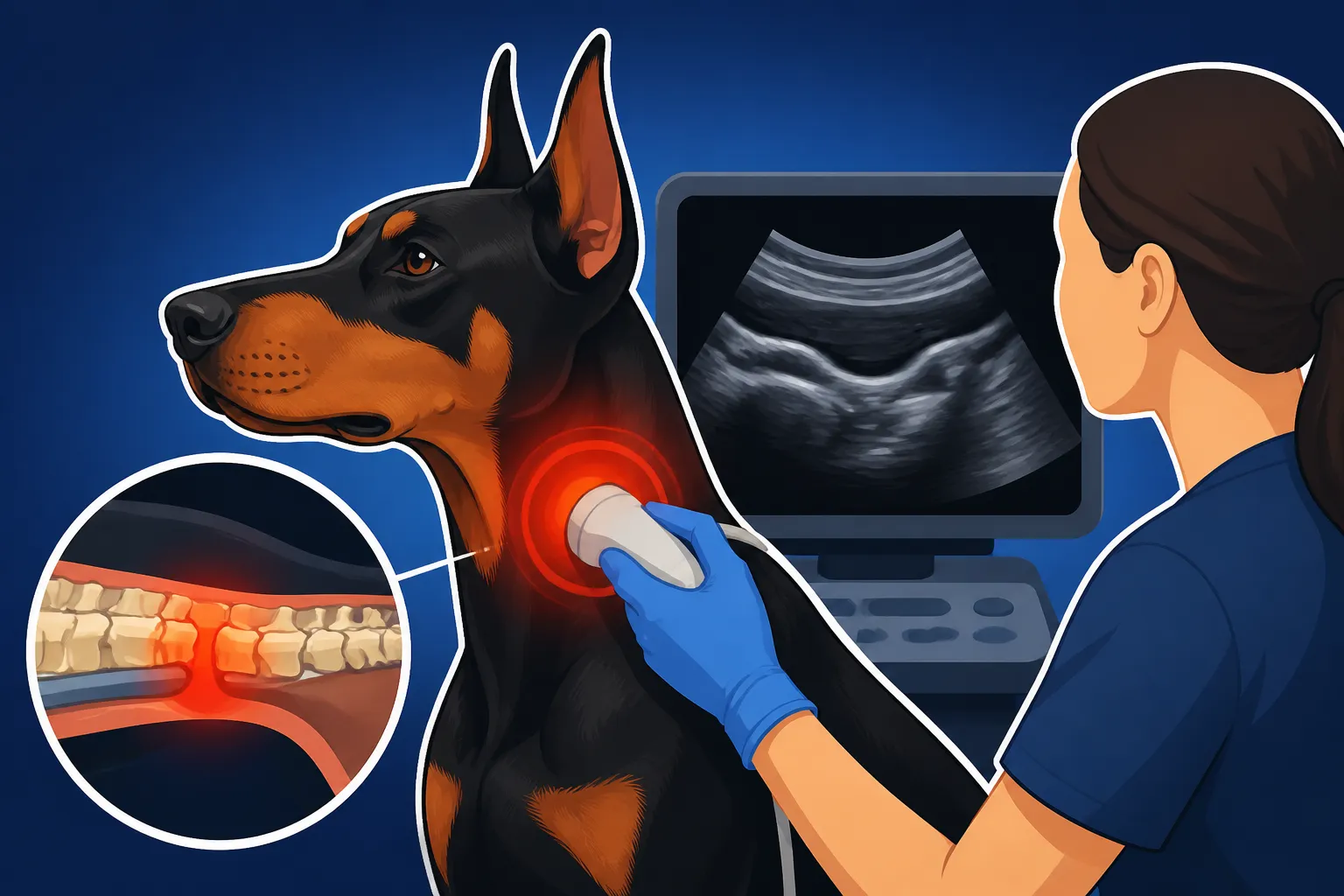
A crucial part of this exam involves neck palpation and range of motion tests. Your vet will gently feel along your Doberman’s neck and spine, looking for tender spots, muscle spasms, or abnormalities. They might slowly extend, flex, and rotate your Doberman’s head and neck to see if any specific positions elicit pain or worsen neurological signs. The goal of this meticulous examination is not just to confirm a problem but to localize it as precisely as possible within the cervical (neck) spine. This localization guides the next steps in diagnostic imaging, helping to narrow down where the compression might be occurring.
Plain Radiographs: Seeing the Bones, Not the Problem
After the neurological exam points towards a cervical spinal issue, plain radiographs, or X-rays, are typically the first imaging modality your vet will recommend. X-rays are non-invasive, relatively quick, and less expensive than advanced imaging. They can provide valuable information about the bony structures of your Doberman's neck.
What can X-rays show? They can reveal obvious vertebral malformations, signs of arthritis in the spine, narrowing of disc spaces between vertebrae, and sometimes, indirect evidence of instability. Your vet might take multiple views, including stress views where the neck is gently positioned in flexion and extension, to try and reveal dynamic compression that only occurs with movement.
However, it’s absolutely critical to understand the limitations of plain X-rays for Wobbler Syndrome. While they're excellent for bone, they do not directly visualize the spinal cord or the soft tissues that often cause compression, such as bulging or herniated intervertebral discs or thickened ligaments. Imagine trying to diagnose a problem with a garden hose by only looking at the concrete path it lies on – you might see an imprint, but you won't see if the hose itself is kinked or blocked. This means that even a Doberman with severe spinal cord compression can have "normal" looking X-rays. Because of this, while X-rays are a good initial screening tool, a definitive diagnosis of Wobbler Syndrome almost always requires more advanced imaging. It helps to understand Why Doberman Neck Vertebrae Compress the Cord which often involves soft tissue issues not seen on plain film.
Unveiling the Hidden Truth: Advanced Imaging Options
When X-rays don't provide a clear answer, or when the neurological signs are severe and progressive, your veterinarian will recommend advanced imaging techniques. These methods allow for a much more detailed view of the spinal cord and surrounding structures, finally revealing the exact nature and location of the compression.
Myelography: The Dye and the Danger
Historically, myelography was considered the gold standard for diagnosing Wobbler Syndrome before the widespread availability of MRI. This procedure involves injecting a radiopaque contrast dye directly into the subarachnoid space around the spinal cord, followed by a series of X-rays or a CT scan. The dye outlines the spinal cord, and any areas where the spinal cord is compressed will appear as a thinning or interruption in the dye column.
Myelography is performed under general anesthesia, and it is an invasive procedure. While it can be effective in visualizing spinal cord compression, it carries certain risks. The most significant risk is the potential for seizures, which can occur as a reaction to the contrast dye, especially in breeds like Dobermans who may already be predisposed to neurological sensitivities. Other risks include hemorrhage at the injection site, worsening of neurological signs, or an allergic reaction to the dye. Because of these risks and the advent of safer, more detailed imaging, myelography is now less commonly performed as a standalone diagnostic, often being integrated with a CT scan for enhanced detail.
Computed Tomography CT: Detailed Bone Views
Computed Tomography, or CT scanning, uses a series of X-rays taken from different angles to create cross-sectional images of the body. These images are then reconstructed by a computer to produce detailed 3D views. CT scans excel at visualizing bone structures with incredible precision, making them excellent for identifying bony malformations, fractures, or significant arthritic changes that might be contributing to spinal cord compression.
For Dobermans with Wobbler Syndrome, a CT scan can clearly show bony spurs, severe degenerative changes in the vertebrae, or instability between vertebral segments. When combined with myelography (CT myelography), it provides a much clearer picture of how bony structures might be impinging on the dye column, indicating spinal cord compression. The benefits of CT include its speed and superior bone detail compared to MRI. However, while it's better than plain X-rays for soft tissue, it still doesn't offer the same level of detail for the spinal cord itself, or for subtle disc herniations and ligamentous hypertrophy, which are frequently the primary culprits in Dobermans.
Magnetic Resonance Imaging MRI: The Ultimate Window to the Spine
For a definitive diagnosis of Wobbler Syndrome in Dobermans, Magnetic Resonance Imaging (MRI) is widely considered the gold standard. MRI technology uses strong magnetic fields and radio waves to generate highly detailed images of soft tissues, including the spinal cord, nerves, intervertebral discs, and ligaments. Unlike X-rays or CT, MRI can directly visualize the spinal cord and precisely pinpoint areas of compression, identify the exact cause (e.g., a bulging disc, a herniated disc, a thickened ligament, or a combination of these), and assess the severity of the spinal cord injury itself.
An MRI provides superior contrast between different soft tissues, allowing veterinary neurologists to see subtle changes that would be missed by other imaging techniques. For instance, it can clearly show if a disc is bulging into the spinal canal, if ligaments are abnormally thick, or if there's inflammation or edema within the spinal cord due to compression. This level of detail is invaluable for guiding surgical planning, as it tells the surgeon exactly what needs to be addressed. Just like myelography and CT, an MRI requires general anesthesia because the patient must remain perfectly still for an extended period. Understanding What a Doberman MRI Shows for Wobblers is critical for both diagnosis and treatment planning.
Navigating the Diagnostic Journey: Preparation and Understanding
Undergoing advanced imaging for your Doberman's neck can feel daunting, especially with the added complexities unique to the breed. Being prepared and understanding what to expect will ease some of that stress.
Anesthesia Protocols and Doberman-Specific Risks
Advanced diagnostic imaging like MRI and CT require your Doberman to be under general anesthesia. This is a significant consideration for any dog, but especially for Dobermans, who are known to have specific health predispositions that can complicate anesthesia. Before any anesthetic procedure, your vet will perform comprehensive pre-anesthetic blood work to assess organ function and ensure your Doberman is as healthy as possible for anesthesia.
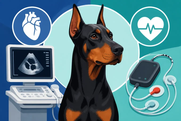
A critical concern for Dobermans is their predisposition to Dilated Cardiomyopathy (DCM), a severe heart condition. Because of this, many vets recommend a cardiac workup, including an echocardiogram, before elective anesthesia. While this might seem like an extra step, it's a vital precaution to identify any underlying heart issues that could increase anesthetic risk. Additionally, Dobermans are known to be predisposed to Von Willebrand Disease (vWD), a common inherited bleeding disorder. While routine blood tests can sometimes flag clotting issues, a specific vWD test may be recommended before surgery or invasive procedures to ensure your Doberman can clot normally. If your Doberman is positive for vWD, special precautions, like administering desmopressin before surgery, may be taken to minimize bleeding risk. Discussing these specific Doberman health concerns with your veterinary team, particularly if you have pet insurance like Healthy Paws, is crucial to ensure the safest possible anesthetic protocol. You can find more information about how this applies to common procedures in Vaccination and Anesthesia in a Doberman.
Decoding the Images: What Neurologists See
Once the MRI or CT scans are complete, the images are sent to a board-certified veterinary neurologist or radiologist for interpretation. These specialists have extensive training in identifying subtle abnormalities and understanding their clinical significance. They will meticulously review hundreds of cross-sectional images, looking for:
- Location of Compression: Pinpointing the exact vertebral level(s) where the spinal cord is being compressed.
- Cause of Compression: Determining if it’s a bulging disc, a herniated disc, a thickened ligament (like the dorsal anular ligament or ligamentum flavum), bone spurs, or a combination. In Dobermans, often multiple factors are at play, sometimes at several spinal levels.
- Severity of Compression: Assessing how much the spinal cord is being squeezed and if there's any evidence of spinal cord damage (e.g., edema or myelomalacia).
- Dynamic vs. Static Compression: Identifying if the compression changes with neck position (dynamic) or is constant (static).
The radiologist will then generate a detailed report, which your primary veterinarian or neurologist will use to explain the findings to you. This report, often accompanied by the actual images, provides the roadmap for treatment decisions, whether conservative management or surgical intervention.
When a Spinal Tap is Necessary
In some cases, even after advanced imaging, your veterinary team might recommend a cerebrospinal fluid (CSF) tap, also known as a spinal tap. This procedure involves collecting a small sample of the fluid that surrounds the brain and spinal cord. It's typically done under general anesthesia, often at the same time as an MRI.
A CSF analysis is usually performed when there's a suspicion of inflammatory, infectious, or cancerous conditions affecting the central nervous system, which can sometimes mimic the signs of Wobbler Syndrome. While Wobbler Syndrome itself is a mechanical compression, a CSF tap can help rule out other concurrent or confounding neurological diseases. By analyzing the cell count, protein levels, and looking for abnormal cells, the CSF analysis provides additional diagnostic information, ensuring that all possible causes for your Doberman’s symptoms are considered and addressed.
The Reality of Costs and Crucial Decisions
Let's be realistic: investigating a complex neurological condition like Wobbler Syndrome in a Doberman is not inexpensive. The financial implications can be a significant source of stress for owners, but understanding the breakdown of costs and the role of pet insurance can help you prepare.
Breaking Down the Bills: What Advanced Scans Cost
The cost of diagnostic imaging for a compressed Doberman neck can vary widely depending on your geographic location, the veterinary specialty hospital, and the specific tests performed.
- Plain Radiographs (X-rays): These are the most affordable initial step, typically ranging from a few hundred dollars.
- Myelography (often combined with CT): This procedure, including the anesthesia, dye, and imaging, can easily run into the thousands of dollars, often between $1,500 and $3,000+.
- Computed Tomography (CT) Scan (without myelography): A standalone CT scan for the cervical spine might range from $1,000 to $2,500+.
- Magnetic Resonance Imaging (MRI) Scan: As the gold standard, MRI is also the most expensive option. For a Doberman's cervical spine, you can expect costs to range from $2,500 to $5,000+, potentially more if other spinal regions are also scanned or if a CSF tap is included.
These figures often include the anesthesia, the scan itself, and the interpretation by a specialist. However, they typically don't cover the initial vet consultation, blood work, or any subsequent treatment. It's a significant financial commitment, and it's important to be aware that these are just the diagnostic costs. Surgical intervention, if recommended, will add many more thousands of dollars to the bill. For a detailed breakdown, you might want to look into What a Doberman Neck MRI Costs.
The Role of Pet Insurance in Covering Wobbler Diagnostics
This is where pet insurance can truly be a lifesaver. Given the high costs associated with diagnosing and treating Wobbler Syndrome, having a robust pet insurance policy can make a world of difference in your ability to pursue the best care for your Doberman without crippling financial strain. Many pet insurance providers, including Healthy Paws, offer policies that cover diagnostic tests, specialist consultations, imaging (like MRI and CT), medications, and even rehabilitation therapies for conditions like Wobbler Syndrome.
However, there's a crucial caveat: pet insurance typically does not cover pre-existing conditions. This means if your Doberman has already shown symptoms of Wobbler Syndrome or received any diagnosis related to a compressed neck before you enroll them, that specific condition will likely be excluded from coverage. This highlights the importance of enrolling your Doberman in a pet insurance plan early in life, ideally before any significant health issues arise. Waiting until symptoms appear is often too late to get coverage for that specific condition. Learning more about Insuring a Doberman Before the Heart Turns and understanding Why a Doberman Insured at Four Is Late can help you make informed decisions about protecting your Doberman’s health and your wallet.
Collaborating with Your Vet for the Best Path Forward
Facing a potential Wobbler diagnosis is incredibly stressful, and the financial burden adds another layer of complexity. It's essential to have open and honest conversations with your veterinary team. Discuss your financial limitations, your Doberman’s quality of life, and the potential outcomes of different diagnostic and treatment pathways. Your vet should be able to provide clear estimates for each stage of the diagnostic process and discuss all available options, including whether less expensive, but less definitive, diagnostics might be appropriate as a first step.
Remember, the goal is to achieve an accurate diagnosis to formulate the most effective treatment plan. While cost is a major factor, an incorrect or incomplete diagnosis can lead to ineffective treatments, prolonged suffering for your Doberman, and ultimately, even greater expense down the road. Working collaboratively with your vet, asking questions, and understanding all your options will help you make the most informed decision for your beloved Doberman.
Beyond Diagnosis: Charting a Course for Recovery
Once a diagnosis of Wobbler Syndrome is confirmed through advanced imaging, the next step is to formulate a treatment plan. This can range from conservative medical management to complex surgical interventions, all aimed at improving your Doberman’s quality of life and alleviating spinal cord compression.
Conservative Management: When Less is More
For Dobermans with mild symptoms, or those where surgery isn't an immediate option, conservative medical management is often the first approach. This involves a multi-modal strategy focused on reducing inflammation, managing pain, and restricting activity to prevent further injury.
- Activity Restriction: This is paramount. Your Doberman will likely need strict crate rest or very limited activity for several weeks, often accompanied by strict leash walks only for bathroom breaks. This helps to reduce stress on the neck and allows any inflammation to subside.
- Medication: Non-steroidal anti-inflammatory drugs (NSAIDs) are commonly prescribed to reduce pain and inflammation. In some cases, muscle relaxants or gabapentin (for neuropathic pain) might also be used. For acute flare-ups, a short course of corticosteroids might be considered to aggressively reduce inflammation around the spinal cord, as discussed in Steroids and Rest for a Doberman Wobbler.
- Physical Rehabilitation: Once initial pain is controlled, physical therapy can play a crucial role. This might include hydrotherapy, therapeutic exercises, massage, and laser therapy to build muscle strength, improve proprioception, and maintain range of motion in the limbs.
- Neck Support: A neck brace or cervical collar may be recommended to limit neck movement and provide support during the recovery phase.
Conservative management can be very effective for some Dobermans, especially those with intermittent or mild signs. However, it's a commitment that requires patience and strict adherence to the vet's instructions.
Surgical Solutions: Decompressing the Spine
For Dobermans with severe, progressive, or intractable neurological signs, surgical intervention is often the most effective way to decompress the spinal cord and improve long-term outcomes. The goal of surgery is to remove the source of compression, whether it's a herniated disc, thickened ligament, or bony abnormality. There are several surgical techniques, and the choice depends on the specific location and nature of the compression identified on the MRI.
Common surgical approaches include:
- Ventral Slot Decompression: This involves approaching the spine from the underside of the neck, removing a small window of bone from the vertebral bodies to access and remove disc material that is compressing the spinal cord.
- Dorsal Laminectomy: This approach involves removing a portion of the vertebral arch from the top (dorsal) side to relieve compression, often used when the compression is on the top or sides of the spinal cord.
- Distraction-Stabilization Techniques: These procedures aim to open up the space around the compressed spinal cord and then stabilize the vertebrae using implants like screws, plates, or bone cement. This can involve replacing a diseased disc with a bone graft and plate (fusion surgery).
Each surgical technique carries its own risks and benefits, and success rates can vary. Post-operative care is extensive, involving strict rest, pain management, and often several months of physical rehabilitation. The decision between surgery and medical management is a complex one, deeply explored in articles like Surgery Against Management for Doberman Wobblers. Owners must be prepared for the significant commitment of time and finances, as Wobbler Surgery Prices for a Doberman can be substantial, and ongoing Rehabilitation Costs After Doberman Spinal Surgery can add up. However, for many Dobermans, successful surgery can lead to a dramatic improvement in their quality of life, allowing them to walk and function much better.
Life After Diagnosis: Management and Prognosis
Regardless of the treatment path chosen, a Doberman diagnosed with Wobbler Syndrome will require lifelong management and monitoring. Even after successful surgery, there's always a possibility of new compression developing at an adjacent site or the progression of degenerative changes elsewhere in the spine. Regular follow-up appointments, including neurological re-evaluations, are crucial.
Owners will need to be vigilant for any recurrence of symptoms and maintain an environment that supports their Doberman's spinal health. This includes avoiding activities that put excessive strain on the neck (like jumping or rough play), using harnesses instead of neck collars, and ensuring they maintain a healthy weight. While Wobbler Syndrome is a serious condition, with an accurate diagnosis and appropriate treatment, many Dobermans can go on to live comfortable and happy lives, even if they never regain 100% of their previous mobility.
| Imaging Technique | What it Shows Best | Pros | Cons | Estimated Cost Range |
|---|---|---|---|---|
| Plain X-rays | Bony structures, gross vertebral alignment | Non-invasive, quick, relatively inexpensive | Does not visualize spinal cord or soft tissue compression | $200 – $500 |
| Myelography | Spinal cord outline, areas of compression | Directly shows spinal cord impingement | Invasive, general anesthesia, risk of seizures | $1,500 – $3,000+ |
| CT Scan | Detailed bony architecture, some soft tissue | Excellent for bone, faster than MRI, good for malformations | Less detailed for spinal cord/soft tissue than MRI, general anesthesia | $1,000 – $2,500+ |
| MRI Scan | Highly detailed soft tissue, spinal cord, discs, ligaments | Gold standard for soft tissue, direct visualization of compression | Most expensive, longer scan time, general anesthesia | $2,500 – $5,000+ |
| CSF Analysis | Inflammatory/infectious markers in spinal fluid | Rules out other neurological diseases | Invasive, requires general anesthesia, separate from imaging | $500 – $1,000 |
Disclaimer
The information provided in this guide is for informational purposes only and does not constitute veterinary advice. Always consult with a qualified veterinarian or veterinary specialist for any health concerns or before making any decisions about your Doberman's medical care.