What a Doberman Echo Actually Measures
It's an incredibly gut-wrenching moment when your vet suggests a cardiac ultrasound, or "echo," for your beloved Doberman. You might feel a knot in your stomach, wondering what exactly they're looking for, what those complex measurements mean, and if this test is truly going to give you the answers you desperately need. This guide is here to demystify the Doberman echo, explaining in plain language what your cardiologist is measuring, what those numbers signify for your dog's heart health, and how this crucial screening tool plays a role in managing a condition like Dilated Cardiomyopathy (DCM).
Understanding the Basics of a Doberman Cardiac Ultrasound (Echo)
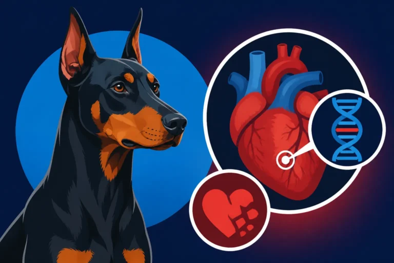
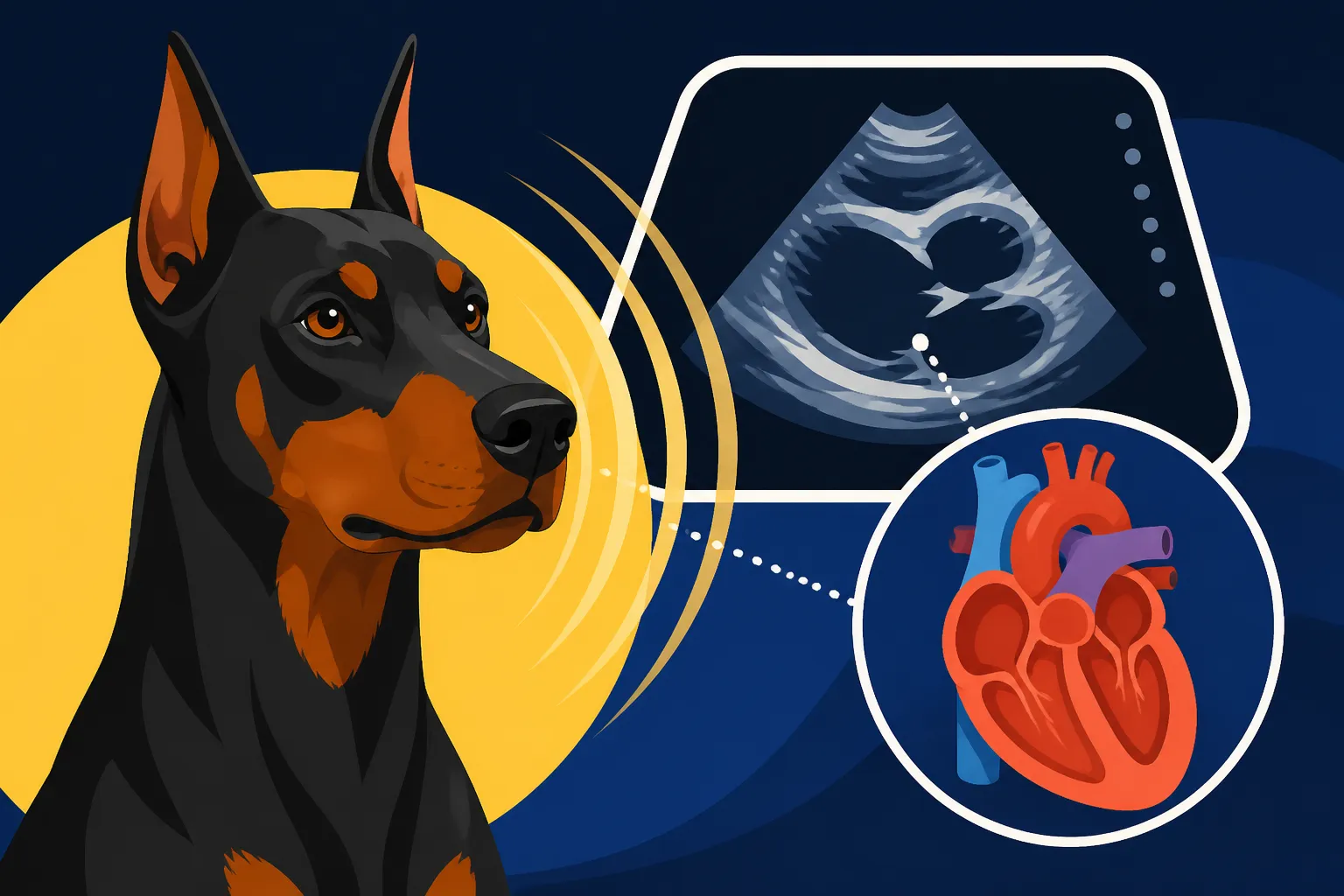
When your Doberman goes in for an echo, it's not just a quick peek at their heart. It's a detailed, non-invasive imaging technique that uses sound waves to create a real-time, moving picture of the heart. Think of it like a sophisticated sonar for the heart, allowing the veterinary cardiologist to visualize its structure, function, and blood flow. For Dobermans, known for their predisposition to Dilated Cardiomyopathy (DCM), this test isn't just routine; it's absolutely vital. DCM is a progressive disease where the heart muscle becomes thin and weakened, leading to an enlarged heart that struggles to pump blood effectively. This can sadly progress silently for years before clinical signs appear, making early detection through an echo incredibly important.
What the Ultrasound Probe Shows the Cardiologist
The cardiologist uses a transducer, which is that small, handheld probe, to send high-frequency sound waves through your Doberman's chest. These sound waves bounce off the heart's structures and return to the transducer, which then translates them into images on a monitor. What they're looking at is a dynamic view of your dog's living, beating heart. They can see the four chambers – the two atria and two ventricles – along with the valves that regulate blood flow, and the major blood vessels entering and exiting the heart. It's truly amazing technology that allows them to assess everything from wall thickness to the integrity of the valves. Without an echo, we'd be relying on more generalized signs like a murmur heard through a stethoscope, which often only flags an issue once it's already well-established. This specific visualization capability is why it's such a cornerstone of preventative care for the breed.
How Your Doberman's Heart Activity is Measured
During the echo, the cardiologist isn't just observing; they're taking precise measurements. They'll use different modes of ultrasound, like 2D, M-mode, and Doppler, to get a comprehensive picture. 2D mode gives a cross-sectional view of the heart, showing the overall shape and size of the chambers. M-mode provides a one-dimensional "ice-pick" view over time, allowing for extremely accurate measurements of chamber dimensions and wall thickness during both contraction (systole) and relaxation (diastole). This is particularly important for detecting the subtle changes indicative of DCM. Doppler ultrasound, on the other hand, allows them to assess blood flow velocity and direction, which helps identify issues like leaky valves or abnormal shunts. All of these measurements combine to provide a detailed report on your Doberman's cardiac health, moving beyond just a visual assessment to hard, quantifiable data.
Key Cardiac Measurements an Echo Provides
The numbers your cardiologist presents after an echo can seem like a foreign language, full of acronyms and percentages. But each one tells a crucial part of the story about your Doberman's heart function. These measurements are compared against established breed-specific reference ranges to determine if there are any deviations that suggest a developing condition, especially Dilated Cardiomyopathy. Understanding these key metrics can help you engage more effectively with your vet and feel more informed about your Doberman's health journey.
Left Ventricular Size and Function
The left ventricle is arguably the most important chamber when it comes to DCM in Dobermans. It's the powerhouse that pumps oxygenated blood out to the rest of the body. In DCM, this ventricle begins to dilate, or enlarge, and its ability to contract effectively diminishes.
- Left Ventricular Internal Diameter (LVIDd and LVIDs): These are crucial M-mode measurements. LVIDd stands for "left ventricular internal diameter in diastole," which is the measurement of the chamber when the heart is relaxed and full of blood. LVIDs is "left ventricular internal diameter in systole," measured when the heart is contracted and pushing blood out. In Dobermans with DCM, both of these values will typically be larger than normal, indicating the chamber is stretched out. A significant increase in LVIDs, especially, tells us the heart isn't contracting as efficiently.
- Fractional Shortening (FS): This percentage tells us how much the left ventricle shrinks during each heartbeat. It's a direct indicator of the heart's pumping strength. A healthy Doberman will have an FS in a certain range, but in DCM, this number drops significantly. It means the heart muscle isn't able to squeeze as effectively, leading to less blood being ejected with each beat. This is one of the most critical indicators of systolic dysfunction, a hallmark of DCM.
- Ejection Fraction (EF): Similar to fractional shortening, ejection fraction is another key measure of the left ventricle's pumping efficiency. It represents the percentage of blood pumped out of the ventricle with each contraction. A lower ejection fraction suggests a weaker heart muscle and less effective pumping, directly aligning with the pathology of DCM. Both FS and EF are invaluable in diagnosing and monitoring the progression of the disease.
Atrial Size and Valve Integrity
While the ventricles often get the most attention with DCM, the atria and heart valves also play critical roles and are thoroughly assessed during a Doberman echo. Problems in these areas can also signal heart disease or contribute to its progression.
- Left Atrial to Aortic Root Ratio (LA:Ao Ratio): This is another standard measurement that cardiologists take. The left atrium receives oxygenated blood from the lungs before it flows into the left ventricle. The aortic root is the beginning of the aorta, the main artery leaving the heart. When the left ventricle starts to fail and blood backs up, the left atrium can enlarge. Comparing its size to the aortic root provides a ratio that can indicate left atrial enlargement, which is a common finding in Dobermans experiencing the early stages of DCM or those with significant mitral valve regurgitation. An elevated LA:Ao ratio suggests increased pressure or volume in the left side of the heart.
- Mitral and Tricuspid Valve Assessment: The heart has four valves that act like one-way doors, ensuring blood flows in the correct direction. The mitral valve is between the left atrium and left ventricle, and the tricuspid valve is between the right atrium and right ventricle. During an echo, the cardiologist carefully examines these valves for any thickening, prolapse (bulging back), or signs of regurgitation (leakage). While DCM is primarily a muscle disease, significant mitral or tricuspid valve regurgitation can sometimes be a secondary effect of the heart's chambers dilating, pulling the valve leaflets apart. Doppler ultrasound helps identify and quantify any such leakage, showing how much blood flows backward with each beat. For some Dobermans, especially as they get older, Why a Doberman Heart Fails Before Anyone Notices can involve these valve issues contributing to overall heart strain.
Wall Thickness and Overall Heart Structure
Beyond just the size of the chambers, the thickness of the heart walls and the overall structure are vital clues to your Doberman's cardiac health. These measurements help differentiate DCM from other types of heart disease and provide insight into how the heart muscle is coping.
- Interventricular Septum (IVS) and Left Ventricular Free Wall (LVFW) Thickness: The interventricular septum is the wall separating the left and right ventricles, and the left ventricular free wall is the outer wall of the left ventricle. In Dobermans with DCM, these walls often become thinner than normal. This thinning is a key characteristic of the disease, as the muscle itself is weakening and stretching. The cardiologist will measure these thicknesses in both diastole (IVSd, LVFWd) and systole (IVSs, LVFWs) using M-mode. Thinner walls, especially in combination with enlarged chambers and poor contractility, are strong indicators of DCM.
- Pericardial Effusion (Fluid Around the Heart): While not directly a measurement of the heart muscle, an echo can easily detect the presence of fluid around the heart, known as pericardial effusion. This isn't a typical finding in uncomplicated DCM, but it can occur in severe cases of heart failure or be indicative of other cardiac problems like tumors. Its presence would prompt further investigation and immediate action, as it can critically impair the heart's ability to fill with blood. Thankfully, for most Dobermans undergoing routine DCM screening, this isn't a concern, but it's part of the comprehensive assessment.
Interpreting Your Doberman's Echo Results
Getting those echo results can feel overwhelming, especially when the vet starts talking about "normal limits" and "borderline changes." It’s important to remember that DCM is a progressive disease, and changes can be subtle initially. An experienced veterinary cardiologist is key to accurately interpreting these findings in the context of your Doberman's breed, age, and overall health. The Age Doberman Heart Disease Usually Starts is often when these subtle changes begin to appear.
What Constitutes a "Normal" Doberman Heart Echo
For a Doberman echo to be considered "normal," all the key measurements – LVIDd, LVIDs, FS, EF, LA:Ao ratio, and wall thicknesses – must fall within established breed-specific reference ranges. These ranges have been determined through extensive research on healthy Dobermans and provide a baseline for comparison. A normal echo indicates that, at the time of the scan, there are no structural or functional abnormalities suggestive of DCM.
However, a "normal" result is a snapshot in time, not a lifetime guarantee. This is particularly true for Dobermans, where DCM can develop later in life. This is why annual screening with an echo and a Holter monitor is so highly recommended. Whether One Clear Echo Is Enough for a Doberman is a question many owners ask, and the answer is typically no; continuous monitoring is essential. Even with clear results, diligent owners know that proactive screening is the best defense against this silent killer.
Recognizing Early Signs of Dilated Cardiomyopathy
The real challenge with Doberman DCM is that the disease has a long "occult phase" – meaning the heart is changing, but there are no outward symptoms. This is why regular echo screening is so vital. Early signs on an echo might include:
- Subtle increases in LVIDd and LVIDs: Even if these aren't drastically out of range, a trend of increasing measurements over successive annual echoes can be a red flag.
- Slight decreases in Fractional Shortening or Ejection Fraction: The heart might still be pumping "adequately," but a drop from previous readings, even if still technically within broad normal limits, can be significant for a Doberman.
- Mild Left Atrial Enlargement (increased LA:Ao ratio): This could indicate the heart is starting to struggle with blood flow, causing pressure to build up in the left atrium.
- Arrhythmias: While primarily detected with a Holter monitor, an echo might sometimes pick up ventricular premature contractions (VPCs) if they occur during the scan. These abnormal heartbeats are often one of the earliest signs of electrical instability in the heart, preceding structural changes.
Catching these subtle changes in The Silent Years Before a Doberman's Heart Shows can allow for early intervention, such as starting medications like Pimobendan, which might slow progression and improve quality of life. This is a topic often discussed by cardiologists, as early treatment has shown promising results in some Dobermans.
What a Cardiologist Looks For Beyond Measurements
While numerical measurements are critical, a skilled cardiologist also uses their experience to observe the overall mechanics and subtle characteristics of your Doberman's heart.
- Regional Wall Motion Abnormalities: Sometimes, one part of the ventricle might be contracting less effectively than another, even if the overall FS or EF is still decent. This localized weakness can be an early indicator.
- Myocardial Texture: The appearance of the heart muscle itself can sometimes offer clues. In some cases, changes in the texture or brightness of the muscle on the ultrasound can be noted, although this is more subjective.
- Dynamic Flow Patterns: Using Doppler, the cardiologist watches blood flow in real-time. Abnormal turbulence, pressure gradients, or subtle regurgitation not captured by a simple measurement can be observed. They are also checking for How a Doberman Heart Enlarges Before Symptoms by seeing how the whole system is responding.
This holistic assessment, combining precise measurements with expert visual interpretation, is what makes the Doberman echo such an invaluable tool for preventing and managing heart disease.
The Importance of Regular Doberman Cardiac Screening
For any Doberman owner, understanding the significance of consistent cardiac screening cannot be overstated. It's not just a vet visit; it's a critical component of responsible Doberman ownership, directly impacting their lifespan and quality of life. The grim reality is that a significant percentage of Dobermans will develop DCM, and it's often a silent progression. That's why being proactive with screening, especially an echo, is the best chance we have to make a difference.
Why Annual Echoes Are Crucial for Dobermans
Given the breed's genetic predisposition to DCM, annual cardiac echoes are absolutely non-negotiable for Dobermans, ideally starting around two to three years of age. The Age to Start Screening a Doberman's Heart is a critical discussion point, but most cardiologists recommend starting early. The disease can progress rapidly, and a heart that appears normal one year could show significant changes the next. Without regular screening, a Doberman could easily progress through the occult phase and into symptomatic heart failure before an owner even notices anything is wrong. This regular check-up allows the cardiologist to establish a baseline for your individual dog and detect those subtle, early changes mentioned above. Early detection buys time, offers more treatment options, and can significantly extend the period of good quality of life for your Doberman. Consider it a yearly health investment, much like you would for yourself. Many Doberman owners rely on pet insurance providers like Healthy Paws to help manage the recurring costs of these crucial screenings and potential long-term medications.
Combining Echo with Holter Monitoring for Comprehensive Screening
While an echo provides an incredible amount of information about the heart's structure and function, it's not the only piece of the puzzle. It's often recommended to combine annual echoes with a 24-hour Holter monitor reading. The Holter monitor is a wearable ECG device that records every single heartbeat over an extended period, typically 24 hours, while your Doberman goes about their normal activities.
| Measurement Tool | What it Measures | Why it's Important for Dobermans |
|---|---|---|
| Echocardiogram (Echo) | Heart structure, chamber size, wall thickness, valve function, pumping efficiency (FS, EF) | Detects structural changes of DCM, assesses severity, guides medication timing |
| Holter Monitor | Heart rhythm, frequency of abnormal beats (VPCs), arrhythmias over 24 hours | Detects electrical abnormalities (arrhythmias) which often precede structural changes in DCM |
The reason for this dual approach is that DCM in Dobermans often manifests first as electrical abnormalities (arrhythmias) before significant structural changes in the heart are visible on an echo. A Doberman with a perfectly normal echo might still have a concerning number of Ventricular Premature Contractions (VPCs) on a Holter. This is why Annual Echo and Holter Screening for a Doberman is the gold standard for comprehensive early detection. The echo tells us how the heart looks and pumps, while the Holter tells us how it's beating. Both pieces of information are vital for a complete picture of your Doberman's heart health.
What Happens After an Abnormal Echo Result
If your Doberman receives an abnormal echo result, particularly one indicative of early DCM, it's a tough pill to swallow. However, it's not a death sentence, especially if caught early. The next steps will depend on the severity of the findings.
- Discussion with your Cardiologist: The cardiologist will explain the findings in detail, discussing which measurements are abnormal and what they signify. They will also outline the prognosis and potential treatment options.
- Medication: For Dobermans in the occult phase (pre-symptomatic), medications like Pimobendan might be recommended. This drug has been shown to improve the heart's pumping efficiency and can potentially delay the onset of clinical signs and extend life expectancy. Other medications, like antiarrhythmics, might be prescribed if significant arrhythmias are present.
- Lifestyle Adjustments: While exercise generally remains important, your cardiologist might advise modifications to your Doberman's activity level, especially avoiding intense, strenuous activity if the heart function is significantly compromised.
- More Frequent Monitoring: Once a Doberman is diagnosed with DCM, whether in the occult or symptomatic phase, monitoring will become more frequent. This might involve echoes every 6-12 months, and regular Holter monitoring. Your cardiologist will tailor a plan specific to your Doberman's needs.
- Ongoing Support: This journey is challenging. Rely on your veterinary team for support and information. Connect with Doberman-specific communities, as there are many owners going through similar experiences who can offer valuable insights and emotional support.
Remember, an early diagnosis from an echo is a gift. It empowers you to take action and give your Doberman the best possible chance at a longer, more comfortable life, even with a challenging condition like DCM.
Disclaimer
This article is intended for informational purposes only and does not constitute medical advice. Always consult with a qualified veterinary professional, especially a board-certified veterinary cardiologist, for any health concerns or before making any decisions about your Doberman's medical care. Every dog is an individual, and only your veterinarian can provide personalized advice based on a thorough examination and diagnostic testing.